Pathological anatomy is a science that studies the pathophysiological and morphological alterations of a disease. That is, it studies the disease at an organic, tissue, cellular and molecular level. For which it uses highly specialized equipment, among them we have the microtome and the cryostat.
What is a microtome?
A microtome is a laboratory equipment that allows the obtaining of thin sections of tissue with a micrometric thickness that can be used later for microscopic study. These equipments are provided with steel, glass or diamond blades, depending on the type of sample to be cut and the thickness in which the tissue sections are needed.
Types of microtomes
Microtomes can be classified into different categories, and each of these will be focused on the specific need to be covered:
- Semi-automatic microtomes
- Automatic microtomes
- Cryostats or Freezing Microtome
- Ultramicrotome
- Slide microtome
- Oscillation microtome
- Universal rotation microtome
- Rotating microtomes
- Manual rotary microtomes
Function of a microtome
Microtomes are equipment that has the function of making very fine cuts to tissues that are generally previously hardened by methods such as freezing, insertion in paraffin or in celloidin. The microtomes have a micrometric wheel that sets the precision and thickness of the blades, which will define the sections or cuts.
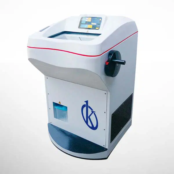
What is a cryostat?
The cryostat is a piece of equipment used in the processing of samples for histological or histopathological diagnosis or identification. It is used mainly to freeze and obtain sections of tissues or frozen samples that have not been previously fixed by chemical methods, to avoid losing some important and necessary characteristics for observation.
This instrument is used in pathological anatomy laboratories in order to obtain tissue sections from frozen material and thus achieve thicknesses ranging from 8 to 40 µm, for analysis under the light microscope.
How does a cryostat work?
The rotation and cut-off mechanism of the cryostat is located inside a refrigerated chamber, at a temperature between -20 and -30 ° C. In this chamber is the sample, the blade and it is where the sections are collected, a process by which the sections are glued to a slide.
Freezing is usually done on a platform within the refrigerated chamber itself with the Peltier system. You can also freeze the tissue externally as quickly as you want, for example with liquid nitrogen, but it is convenient to place the sample in the cryostat chamber until its temperature is equal to that of the cryostat to obtain homogeneous sections. Before freezing, the sample is embedded in a medium that is liquid at room temperature and solid at cut-off. Therefore, we have our sample in a solid block, embedded but not included. This allows the sample to be manipulated and attached to a sample holder, which will be fixed to a shaft that advances on the blade.
At Kalstein we are MANUFACTURERS and we have a wide range of new microtomes and cryostats at the best PRICES. That is why we invite you to take a look at the Products menu. HERE