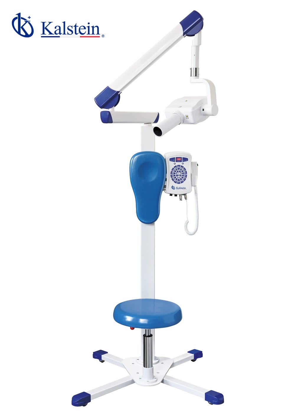
Interpreting Results and Reading Measurements with the YR06196 Digital Veterinary Intraoral X-ray System
The YR06196 Digital Veterinary Intraoral X-ray System represents a significant advancement in veterinary diagnostic imaging technology. This article focuses on the interpretation of results and reading measurements from this sophisticated system, ensuring that users maximize its capabilities for precise diagnostics in veterinary practice.
Understanding the YR06196 Digital X-ray Technology
The YR06196 utilizes advanced international technology with effective integrated design that ensures high efficiency and minimal radiation exposure. This system is particularly well-suited for veterinary practitioners who require accurate imaging for diagnostics. The intelligent microcomputer control not only allows remote exposure control but also includes enhanced low voltage alarm functions and high voltage protection.
The design choices made in the YR06196, such as micro-focus technology, result in sharper imaging and more accurate diagnostics. With two operational modes (mobile and fixed) and a pneumatic lift seat, it caters to both the dentist's convenience and patient comfort.
Technical Data Table of YR06196
| Specification | Detail |
|---|---|
| Model | YR06196 |
| Power Supply | 220V±10%, 50HZ, 1KVA |
| Tube Voltage | 70 kVp |
| Tube Current | 8 mA |
| Focus Size | 0.8 mm |
| Total Filtration | 2.5 mmAL |
| Exposure Time | 0.2-4s |
| Leak Radiation | Outside one meter ≤ 0.002Mg/h (National standard: 0.25Mg/h) |
| Net Weight | 57 kg |
| Pixel Size | 18.5µm |
| Resolution | 14-20 Ip/mm |
| Power Consumption | 600 mW |
Interpreting X-ray Results from YR06196
Accurate interpretation of x-ray results is essential for effective veterinary diagnostics. The YR06196 produces high-quality images that allow veterinarians to identify conditions affecting dental health in animals. Understanding the parameters displayed, including density, clarity, and contrast, is crucial for making sound clinical decisions.
When evaluating x-ray images, practitioners should look for common indicators of dental issues such as periodontal disease, tooth fractures, or abscesses. Familiarity with normal anatomy is essential, as deviations from typical appearances can indicate pathology.
Common Misinterpretations and How to Avoid Them
Misinterpretation of x-ray images can lead to incorrect diagnoses and treatments. Some common pitfalls include mistaking normal anatomical variations for pathological conditions. Training and experience are critical in minimizing these errors.
To improve interpretation, veterinarians should have a systematic approach, including checking for image quality, ensuring optimal exposure settings, and regularly reviewing clinical findings in conjunction with imaging results.
Case Studies: Worked Examples of Image Interpretation
Let's examine a few case studies that highlight the effective interpretation of results obtained from the YR06196. By reviewing actual x-ray images, veterinarians can see the nuances in dental anatomy and how specific findings correlate with clinical presentations.
For instance, a case of an impacted tooth may be clearly visible on the x-ray, showing a dark area surrounding the tooth root indicative of infection. Conversely, a healthy tooth will exhibit a clear outline without such shadows.
Essential Equipment Setup for Accurate Measurements
Proper setup of the YR06196 is essential for obtaining accurate measurements. Unboxing the equipment, anchoring it securely, and ensuring it is level are critical first steps. Once set up, a power-up sequence must be followed, including a first-run calibration to ensure the device functions correctly.
Misalignment or incorrect setup can lead to distorted images, which in turn affects interpretation. A checklist can help ensure all steps are followed correctly to prevent initial setup errors.
Frequently Asked Questions
What are the key features of the YR06196 that aid in accurate interpretation of results?
The YR06196 features advanced image processing technology, a micro-focus capability for sharper images, and customizable exposure settings that enhance diagnostic accuracy. These features collectively help in producing clearer images for better interpretation.
How does the low radiation exposure benefit both the veterinarian and the patient?
Low radiation exposure minimizes risks for both the veterinarian and the patient. It allows for frequent imaging without the cumulative risks associated with higher doses, making it safer for animals undergoing dental evaluations.
What common errors can occur when interpreting x-ray images?
Common errors include misidentifying normal variants as pathological conditions, overlooking subtle changes that indicate disease, and failing to recognize overlapping structures that can obscure key findings. Continuous education and practical experience help mitigate these issues.
How can veterinarians enhance their skills in reading x-ray results?
Regular training sessions, peer reviews, and utilizing digital resources that provide examples of both normal and abnormal findings are effective ways to enhance skills in reading x-ray results. Engaging with case studies can also provide valuable insights.
What maintenance practices ensure the YR06196 remains reliable for accurate diagnostics?
Regular maintenance practices include routine calibration, software updates, and cleaning of components to prevent dust accumulation. Furthermore, regular checks on power supply and connections can ensure consistent performance.
How important is it to have knowledge of normal anatomy when interpreting x-ray results?
Knowledge of normal anatomy is critical. Understanding the baseline anatomical structures allows veterinarians to identify pathological changes accurately. This foundational knowledge leads to improved diagnostic capabilities and patient outcomes.
Can the YR06196 be integrated with other dental imaging systems?
Yes, the YR06196 can connect with various digital dental imaging systems, enhancing a veterinarian's ability to perform comprehensive diagnostics and streamline workflows within the clinic.
If you are looking for a fusion of innovation and quality, you have come to the right place. At Kalstein, we offer you the luxury of exploring our exclusive catalog of laboratory equipment. We manufacture every device to the highest standards of excellence. Our intuitive and seamless online purchasing channels are designed for your convenience, securing the most competitive prices. Hesitate no longer — we bring science to life, it is time to become part of our community.