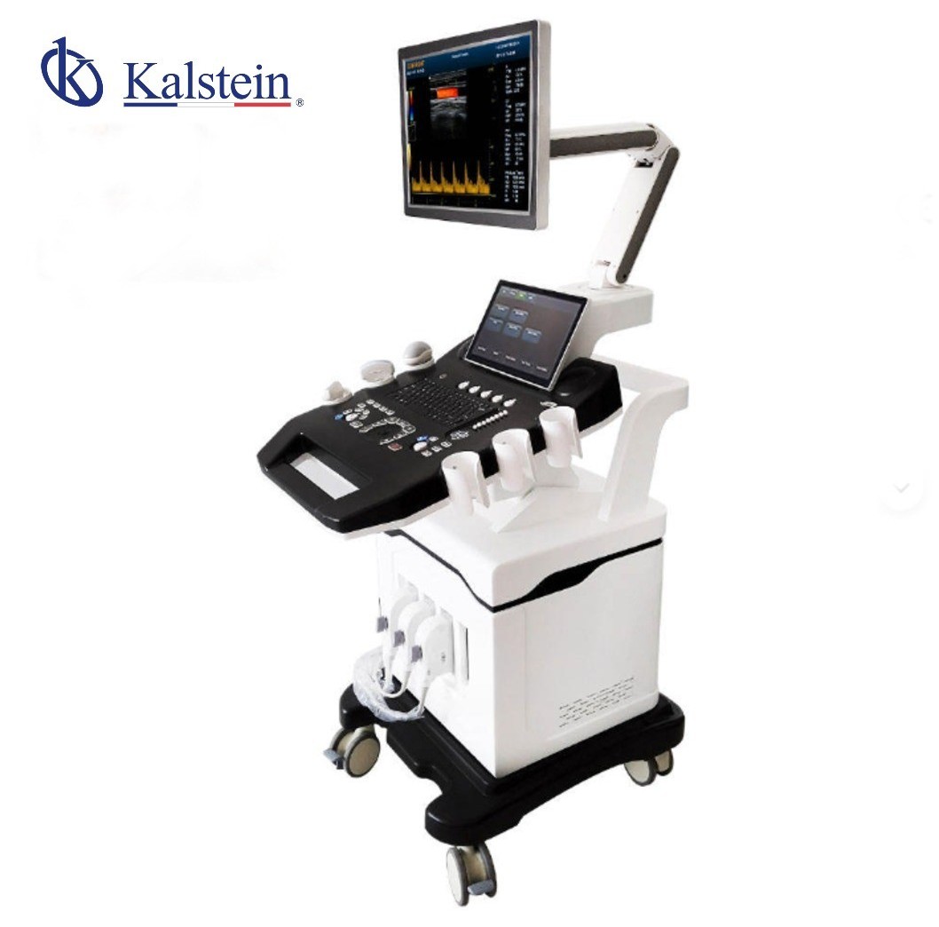
Understanding the Principles and Scientific Foundations of the YR06635 Ultrasound Scanner
The YR06635 is a high-end diagnostic imaging solution that incorporates advanced 3D and 4D Color Doppler technology. This system is tailored for modern medical and veterinary centers that demand exceptional precision across a wide range of applications, including abdominal and cardiac studies, as well as complex obstetric and gynecological imaging.
Scientific Principles Behind the YR06635
The YR06635 operates on the fundamental principles of ultrasound technology, primarily utilizing sound waves to create images of internal body structures. The system employs high-frequency sound waves (between 1 and 40 MHz) that are transmitted into the body via a transducer. When these sound waves encounter tissue, they are reflected back to the transducer, allowing for the creation of high-resolution images on the monitor.
In Doppler ultrasound, especially utilized in the YR06635, the principle of frequency shift is employed. When sound waves are reflected off moving structures, such as blood cells, their frequency changes. This change is measured, allowing for the assessment of blood flow velocity, direction, and volume. The incorporation of both 3D and 4D imaging provides an invaluable advantage, allowing clinicians to visualize structures in real-time and make accurate diagnoses.
Design and Intelligent Technology of the YR06635
The YR06635 incorporates a suite of intelligent technologies that enhance its usability and operational efficiency. It features four active transducer ports and four USB ports, facilitating seamless probe switching and quick data export. The fully digital signal processing technology ensures high-resolution images with reduced noise and artifacts.
Moreover, the advanced visualization capabilities of the YR06635 are impressive. It is equipped with a 15-inch LED monitor featuring a contrast ratio of 800:1 and a swivel angle of ±90 degrees for ergonomic operation. This design consideration is critical in a clinical setting, where quick and accurate assessments are paramount.
Technical Specifications of the YR06635
| Features | Specifications |
|---|---|
| Model | YR06635 |
| Monitor | 15-inch LED (1024 x 768) |
| Transducer Ports | 4 Active Ports |
| Storage | 64GB SSD (Optional 120GB/200GB) |
| USB Ports | 4 Ports |
| Cine Memory | 1200 Frames |
| Image Formats | PNG, AVI, BMP, JPEG, DICOM |
| Imaging Modes | B, 2B, 4B, M, B/M, B/C, PW, CW, CFM, 4D |
| Connectivity | Ethernet (100Mb/1000Mb), VGA, HDMI, DICOM 3.0 |
| Beamformer Frequency | 1 to 40 MHz |
| Equipment Dimensions | 73 x 113 x 144 cm |
| Equipment Weight | 80 kg |
| Package Weight | 200 kg |
Frequently Asked Questions
What types of transducers are compatible with the YR06635?
The YR06635 supports a wide variety of transducers, including convex array, linear array, intracavitary, micro-convex, cardiac, and the specialized 4D volume probe.
Is the YR06635 compatible with hospital management systems?
Yes, it fully complies with DICOM 3.0 protocol, allowing for easy reporting and sending of images through medical networks.
Does the YR06635 offer automated functions?
Yes, it features automatic measurements of IMT (Intima-Media Thickness) and automatic Pulsed Wave (PW) tracing to expedite the diagnostic process.
What are the key advantages of using the YR06635?
The YR06635 is highly versatile, applicable in cardiology, urology, pediatrics, musculoskeletal imaging, and more. It offers complete imaging modes and has an easy-to-use interface based on Windows.
What are some potential disadvantages of the YR06635?
Some considerations include its significant weight of 80 kg, which requires a stable surface or its own internal transport system, and the learning curve related to its high-end nature and multiple imaging modes.
What are the ideal settings for using the YR06635?
The YR06635 is best utilized in high-complexity hospitals and specialized clinics focused on vascular diagnostics or advanced fetal monitoring. The integrated 4D mode is essential for real-time volumetric reconstructions in obstetrics.
How can I request a quote for the YR06635?
You can request a quote for the YR06635 by contacting our sales team directly through our website or inquiry form.
If you are looking for a fusion of innovation and quality, you have come to the right place. At Kalstein, we offer you the luxury of exploring our exclusive catalog of laboratory equipment. We manufacture every device to the highest standards of excellence. Our intuitive and seamless online purchasing channels are designed for your convenience, securing the most competitive prices. Hesitate no longer — we bring science to life, it is time to become part of our community.