An ultrasound scanner is a medical device that provides images of most soft tissues through the use of ultrasonic waves. That is, it allows obtaining diagnostic
An ultrasound scanner is a medical device that provides images of most soft tissues through the use of ultrasonic waves. That is, it allows obtaining diagnostic images from the echoes obtained by the emission of ultrasound waves. These waves are produced by an instrument called a transducer, which, in addition to generating the ultrasound waves, is also capable of detecting the echoes reflected by the ultrasound, generating two-dimensional images of tissues and organs.
How does an ultrasound scanner work?
The transducer picks up the echo from the sound waves and a computer converts it into an image that appears on the screen. In most cases, the elements that make up the transducer are made of ceramic glass called piezoelectric. These are capable of producing sound waves when an electric field passes through them, and they are also capable of working in the reverse direction, producing an electric field when receiving sound waves.
Ultrasounds are high frequency sound waves (more than 20,000 cycles per second or 20 kHz) that are not audible to humans. Different tissues alter the waves in different ways, some reflect it directly and others scatter them as echoes before they reach the transducer. That is, using the speed of sound and the time each echo returns, the scanner calculates the distance between the transducer and the tissue boundary. Deeper reflected echoes are more attenuated than shallow ones. When the echoes return to the transducer it is possible to reconstruct a two-dimensional map of the tissues and organs.
How is an ultrasound scanner composed?
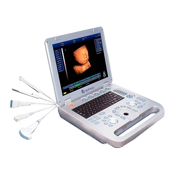
A typical ultrasonic scanner system is composed of a beamformer, a central processing unit, a user interface (keyboard, control panel, trackball), several probes (transducers or scan heads), one or more video displays , some kind of recording device, and a power supply system.
For ultrasonic imaging, a probe is placed on the skin (after applying an acoustic coupling gel) or inserted into a body cavity. As mentioned, ultrasonic probes have elements made of piezoelectric materials (materials that convert electrical energy into acoustic energy and vice versa). When the ultrasonic energy emitted from the probe is reflected by the tissue, the transducer receives some of these reflections (echoes) and transforms them back into electrical signals. These signals are processed and transformed into an image
What are the medical applications of ultrasound scanner?
Ultrasound scanners are used in the radiology department and other clinical departments, as well as in independent imaging centers and private medical practices, primarily for vascular and gynecological-obstetric applications. Some systems include additional transducers to facilitate more specialized diagnostic procedures, such as cardiac, vascular, endovaginal, endorectal, or small part (eg, thyroid, breast, scrotum, prostate) scanning.
At
Kalstein we are MANUFACTURERS of high quality medical equipment at the best PRICES on the market, that is why we present you a new ultrasound scanner and invite you to take a look at the Products menu.
HERE