Electrocardiographs are medical equipment that perform essential work for the diagnosis of heart disease and the diagnosis of heart disease and its cavities.
This device captures and expands all the electrical activity that the heart has, by means of electrodes that are placed in the four (4) extremities of the body and six (6) that go in precordial positions, in the thorax, which records data called electrocardiograms. By means of electrocardiographs, it is possible to discover if the patient has any heart disease, which can compromise his health since the heart is responsible for pumping blood throughout the body.
That is why doctors can detect arrhythmias, blockage of the arteries, presented with symptoms such as chest pain, difficulty breathing well, tachycardias, fatigue, fainting, among others.
Types of the Electrocardiographs
- Single-channel electrocardiograph: They record and print the results of the physical activity of the heart, which specialists must order 12 manual shunts to perform the analysis and has as advantage that it is light and its use is quite useful and simple.
- Multichannel electrocardiograph: this one is characterized by having 3, 6 and 12 channels, which allow to differentiate the time to choose the formats and with the display of screens, in addition to being able to visualize the analysis in real time before generating the impression, facilitating and improving the accuracy of the patient’s diagnosis.
- Printed multichannel: this system has recognition patterns that identify normal and abnormal signals from the results of an electrocardiogram, where the user must verify, complete and study in depth to generate an accurate diagnosis for the patient.
Different studies with the use of electrocardiographs
- Resting electrocardiographs: performed during a consultation or before an operation. It is essential and allows the staff in charge to monitor the general state of health of the patient. It is performed by connecting the electrodes, on the skin, previously shaved and clean, at different points of the body (torso, wrist, ankle). They’ll capture cardiac electrical activity. A minimum of 6 should be used, but it is common to use 12, 15 or even 18.
- Electrocardiograph for stress tests: this is developed during physical activity in patients suffering from palpitations or chest pains. It is usually performed on a running machine, gradually increasing speed, in addition they can measure respiratory rate and blood pressure.
Electrocardiographs en KALSTEIN
As we just mentioned the operation of the electrocardiograph, as a clinical diagnostic equipment, is based on the installation of a series of electrodes on the surface of the patient’s skin at the level of the thoracic region and extremities. These electrodes allow the electrical signal generated by the activity of the patient’s heart to be captured.
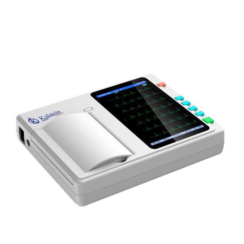
An electrocardiograph is used to measure any damage to the heart, if there are abnormal heart palpitations, the size and position of the heart’s chambers, and effects of drugs or devices used to control the heart (such as pacemakers). Nowadays there are multiple types of electrocardiographs, the simplest ones draw the tracing on a paper tape specific registry for ECG and the most modern and complex ones make an analysis of what they record and store it in digital format. There are 3.6 and 12 channels, which are YR models, which will give them the power of advanced and easy-to-use technologies, multiple forms of data management, as well as accurate measurements and interpretations, which can improve the diagnostic confidence of cardiologists.have 7″ touch screen, 12.5mm/s paper speed, 25mm/s,50mm/s, with recording mode via a thermal printing system, continuous paper, with a floating input circuit, circuit of protection against defibrillator effect, among other specifications that you will find through our link HERE
