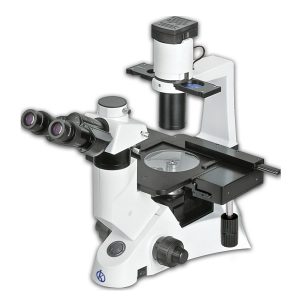
Initially, the use of the field ion microscope, are used for studies of atoms and their external behaviors. It is represented as a tool within the laboratories, which is responsible for analyzing the surfaces of materials, in order to obtain a wide panorama of atomic structures, and to be able to visualize individual atoms. Among its functions, it is able to show the organization of atoms, which are formed on the surface of a metal tip established for such function. And thanks to a magnetic field, developed by certain chemical elements like neon.
For this reason, the field ion microscopy was developed using atomic probe tomography. It is a more advanced technique, which can perceive the three-dimensional atomic structure, and identify to which chemical element the atoms of the samples belong.
Field Ion Microscope Structures
It has a tube on top called a pump, then two tungsten tubes follow, on the outside, a few centimeters from the pump, it has a Board of Indium. Inside the microscope, there is a chamber where solid nitrogen is stored, and two smaller chambers on the sides that contain liquid nitrogen. Below the solid nitrogen chamber, it has a tungsten filament, composed of a test piece. The chamber, filament and test piece are surrounded by a copper cylinder.
At the bottom, at one end, it has a pipe leading to a vacuum pump, and at the opposite end it has a pipe leading to the purification siphon and helium bottle. On the lower deck, where this microscope is almost completed, is a fluorescent screen, supported by an indium seal, which allows the image of the individual atoms.
Development of the Field Ion Microscope
This microscope has great dimensions because of the number of pieces, and the processes it carries out. This device works as follows:
First, a sharp-pointed metal needle is placed in an ultra-high-vacuum chamber, then this chamber is filled with helium or neon, which serve as a visualizing gas. The needle must then be cooled to cryogenic temperatures, i.e. below 100 degrees Celsius. Subsequently, 5000 to 10000 volts of positive voltage are applied to the needle tip. The gas atoms that are assumed by the needle to be ionized by electrons that do a tunnel effect, thanks to the electric field that occurs at the moment, are very close to them.
Deviation of the surface near the tip of the metal needle causes natural magnetization, and what is known as a point projection effect occurs, where ions are sharply resisted, projecting them toward a perpendicular area of the surface, also known as point projection.
Field Ion Microscope Application
This microscope is used to examine the behavior of surfaces, and atoms that live in it. It is commonly used to inquire about planes composed of crystals, to assimilate their dynamics and interactions with other elements.
Also, it is applied to experiment on adhesion phenomena that occur when ions, atoms, gases, molecules, liquid or solid elements, are stagnant on a surface; causing desorption effects, process by which, turns out to be contrary and surface diffusion clusters of adatoms, which are atoms occupying a space on glass surfaces, and the interactions of the adatoms, towards the equilibrium between atoms on a surface, among others.
Kalstein brand microscope
At Kalstein, we offer the best laboratory equipment, among them are the Microscopes, belonging to the YR models, with very attractive general features, such as; 3 W S-LED (LCD screen amplification), suspension timer, brightness lock and indication, etc., Two wave range (B, G, U, V can be combined). Lens lighting fly-eye. application software. Tilt binocular display head. Fivefold backward (no coding). Infinite Optical System, Infinity Siedentopf Binocular Display Head; inclined to 45° interpupillary 47-78 mm. Infinite Phase Contrast Lens. Even modern design lighting, Kohler illumination and aspherical collectors can be adapted. Portable aluminum case available for high level packaging requirements. HERE
We are manufacturers and we have the best advice, so that your purchase is the ideal and at excellent prices. For more information, visit our website HERE


