It is a microscope that allows us to observe conventional images and contrasts with fluorescence, it is worth noting that a traditional optical microscope is used, but with small specifications that allow the optical system to adapt by fluorescence; this feature is called “EPI fluorescence” because it is inserted above the optical system or magnification targets, working by light reflection.
The conventional microscope must have certain characteristics for adaptation to be possible, otherwise when adaptation is required it may not be feasible; to avoid this do not hesitate to consult our range of microscopes through the following link HERE our advisors will attend you; accessories requiring a fluorescence PPE consist of an electric power supply, a light socket including a mercury vapor lamp, a light collecting system and specific fluorescence filters.
Advantages of fluorescence microscopy
At the beginning of the fluorescence microscopy the revolution in cell biology began, this allowed to observe the images of living cells, multiple markings of individual organelles and macromolecular complexes through synthetic and genetically encoded fluorescent probes; using this technique we can obtain images of the distribution of a single molecular species.
Fluorescence microscopy extends the scope of application of the microscopy field, using it to control the location of intracellular components marked with specific fluorophores. As well as their associated diffusion coefficients, transport characteristics and interaction with other biomolecules. Also to observe the localized environmental variables, it allows to investigate the pH, viscosity, refractive index; the ionic concentrations, membrane potential and polarity of the solvent in living cells and tissues.
How a fluorescence microscope works
The first thing is to prepare the sample with a fluorescent substance called fluorophore, then it will be illuminated through the lens with a high energy light. The light will be absorbed by the fluorophore and will cause light emission with low energy and a long wavelength. This fluorescent light can be separated from the surrounding radiation using filters designed for that specific wavelength, allowing the observer to visualize only that which is fluorescent.
The dichroic beam splitter and excitation and emission filters are selected in such a way that the spectral excitation is compatible with the emission characteristics of the fluorophore used to mark the samples and this is how the distribution of a single color or fluorophore is detected at that time. To view multicolored images, you must combine multiple images in a single color.
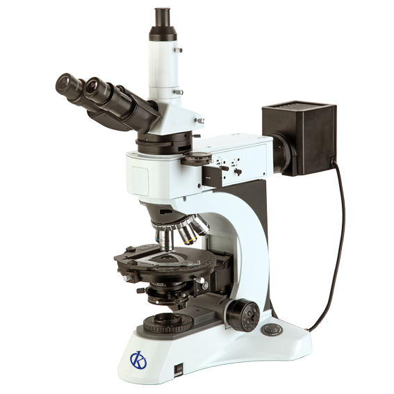
Part of the fluorescence microscope
- An excitation filter.
- A dichroic mirror or beam splitter.
- An emission filter.
- A light source (which can be a xenon arc lamp, a mercury vapor lamp, high power LEDs or lasers).
- A set of lens lenses.
- An eye lens.
- A scenario to contain the sample.
- A detector.
Disadvantages of fluorescence microscopy
The photobleaching resulting from fluorophore illumination can seriously affect the time period of observation of a sample by fluorescence microscopy. This technique used in living cells is prone to increase their toxicity, i.e., fluorescent molecule can produce specific reactive agents when illuminated, further increasing the phototoxic effect.
Although, through fluorescence, a tissue sample with a fluorescent DNA stain could be observed. This could show the organization of DNA within cells, but it cannot reveal anything about cell morphology.
If you want to know the catalog of high-end products that we Kalstein have for you, visit us HERE we have a specialized team that will answer your doubts, remember that choosing the best and most updated microscope is the basis to be able to convert your conventional microscope into one of fluorescence, in addition we assure that through our online sales channels is very easy and viable, remember we are Company manufacturer of Laboratory Equipment recognized worldwide.
