Description
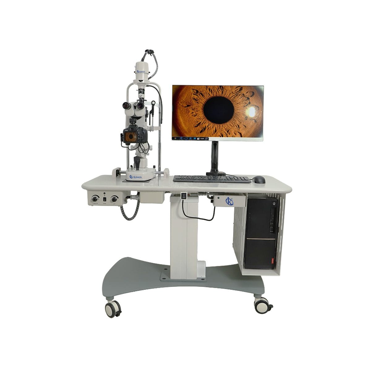
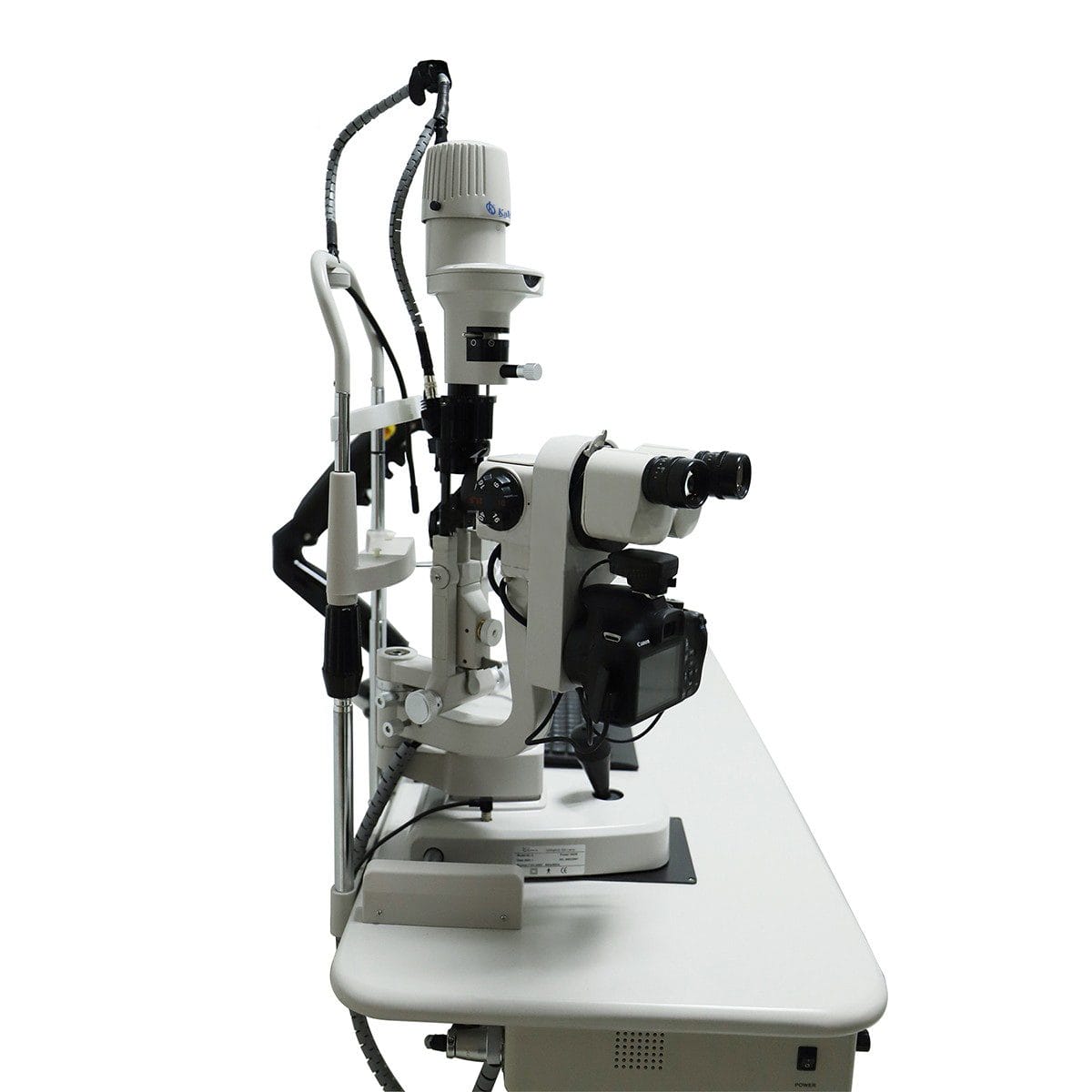
The Veterinary Digital Slit Lamp YR06200 is an innovative tool in the field of veterinary diagnostics, providing an advanced color digital image acquisition system. Its ability to deliver digital images with a resolution greater than 2000 lines ensures exceptional image clarity. With unique case storage capabilities, professionals can store and review real-time images, preserving them permanently, which is essential for thorough examination and record-keeping. The device’s powerful image analysis and processing functions allow for detailed lesion analysis, measurement, and image enhancement, facilitating reliable comparative analysis of therapeutic effects.
Market Price
Digital slit lamps like the Veterinary Digital Slit Lamp YR06200 are generally found within a market price range of $13,000 to $14,500. This range provides potential buyers with an understanding of the competitive landscape, ensuring informed purchase decisions.
Frequently Asked Questions
What are the primary uses of the Veterinary Digital Slit Lamp YR06200? The lamp is primarily used for detailed eye examinations in veterinary settings, enabling precise analysis of ailments.
Is the device portable? While it is designed for clinical use, the portability depends on the specific setup and additional transportation accessories.
Can images be printed directly from the device? Yes, it is equipped with a professional color image inkjet printer for high-accuracy outputs.
Advantages and Disadvantages
Advantages: High-resolution imaging, comprehensive image storage, and advanced analysis capabilities are significant benefits. The professional-grade optic interfaces allow for precise diagnostics.
Disadvantages: The complexity of the device requires trained staff for optimal operation, and the initial investment may be high for small veterinary practices.
Product Usage in Field
In practical terms, the Veterinary Digital Slit Lamp YR06200 is used in veterinary clinics for the detailed examination of animal eyes. Its precision optics and digital processing capabilities make it particularly useful for diagnosing conditions such as cataracts or retinal issues.
Recommendations
To maximize the use of the Veterinary Digital Slit Lamp YR06200, ensure periodic maintenance checks and cleaning of lenses for optimal performance. Training staff in its advanced features will allow you to leverage its full potential for detailed diagnostics.
Features
- Digital image resolution greater than 2000 lines
- Advanced storage capabilities for permanent image preservation
- Powerful lesion analysis and image processing functions
- Equipped with Galilean optical interfaces
- Professional color image inkjet printer included
Technical Specifications
| Model | YR06200 |
| Microscope Type | Parallel angle type |
| Eyepiece | 12.5X |
| Total magnification microscope | 6X , 10X , 16X , 25X , 40X |
| Visual field diameter (mm) | φ37 , φ23 , Ø14 , Ø8 .7 , size 5.7 |
| Diopter adjustment | -5D ~ +5 D |
| Fissure width / height width | 0mm ~ 10mm continuously adjustable, high- 1mm ~ 10 continuously adjustable |
| Spot diameter (mm) | Ø10 , Ø8 , Ø5 , Ø3 , Phi] 2 , φ0.2 |
| Crack angle | 0 ° ~ 180 ° continuously adjustable |
| Fissure angle | 5°, 10°, 15°, 20° |
| Lighting imported | From Germany tungsten 12V/60Hz |
| Magnification | 0.794X (± 8%) |
| Filters insulation | Film, minus rays, no red tablets, cobalt blue chip |
| Input voltage | 110V/220V (± 10%) 50/60Hz |
| Input Power | 60VA |
| Input voltage tungsten halogen | 4.5V , 6.0V , 9.0V , 10.5V , 12V |
| Fixation lamp | 3.5V |


